Hidden Content
Hidden content
Hidden Content
Hidden content
BLOG/
Skin Cancer Disguised as a Mole? Doctor Teaches You How to Identify It at a Glance!
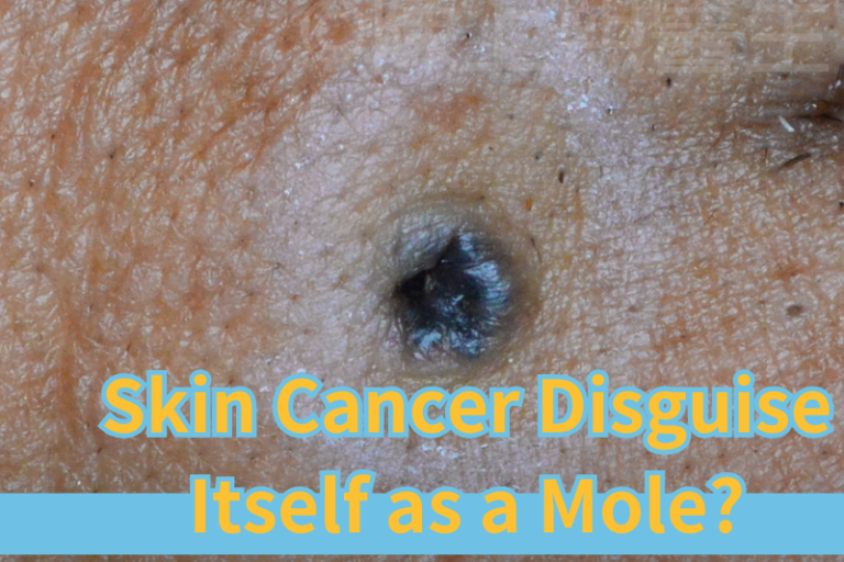
Today, I want to share an interesting and important case. This case involves a male patient approximately 60 years old, who noticed that a mole at the corner of his right mouth was gradually getting larger, and there was a small hole in the center of the mole. He felt uneasy and worried that this mole could be threatening, so he came to the dermatology department for a professional evaluation.
Initial Examination Results
After a detailed examination, we found that the mole exhibited the following characteristics:
- Significantly increased size
- Very smooth surface, resembling a pearl
- Central depression, forming an ulcer
These features are highly consistent with the appearance of pigmented basal cell carcinoma. Basal cell carcinoma is a common type of skin cancer that, although it is less malignant, can still cause local tissue destruction if not addressed promptly, potentially leading to more severe consequences.
Importance of Mole Excision
When a doctor suspects that a mole may be basal cell carcinoma, they typically recommend that the entire mole be completely excised and sent for pathological examination to confirm the diagnosis.
Why Don’t We Use Laser Removal?
Laser removal only targets surface tissues and cannot ensure that deep cancer cells are fully removed from the skin. If cancer cells are not completely eliminated, it may lead to delays in treatment or recurrence of the disease. Therefore, surgical excision is the safest and most thorough treatment method.
How to Identify Basal Cell Carcinoma?
The appearance of basal cell carcinoma can be similar to that of ordinary moles, spots, or wounds, making it easy to overlook. However, it has some typical features:
1.Smooth or pearl-like surface
- The cancerous area typically has a smooth, even reflective surface resembling a pearl.
2.Central depression or ulcer
- The cancerous area may have a central depression or ulcer that does not heal easily.
3.Clear, glossy borders
- The edges of the tumor often appear wavy and reflective.
4.Vascular dilation
- The cancerous area may be accompanied by noticeable vascular dilation, forming a spider web-like pattern of red lines.
If you notice any of these abnormal features on your skin, it is advisable to seek a diagnosis from a professional dermatologist immediately.
Surgical Process Analysis
In the treatment of this patient, we performed a complete excision of the mole under local anesthesia. Here are the key steps of the surgery:
1.Local Anesthesia
- An anesthetic needle is used to numb the skin around the mole, ensuring that the patient feels no pain during the procedure.
2.Complete Mole Excision
- The depth of excision must reach the dermis to ensure that all potential cancer cells are removed. The incision may even reach the subcutaneous fat, which typically appears as yellow tissue.
3.Wound Management and Suturing
- After excision, we need to perform subcutaneous tissue loosening around the wound. This allows the skin on both sides of the wound to be pulled together, reducing tension during suturing.
- Reason for Reducing Tension: If the tension on the wound is too great, it may lead to thick scars or even poor healing or opening of the wound.
4.Sample Submission for Examination
- The excised mole is sent to the pathology department for microscopic analysis to confirm whether it is basal cell carcinoma.
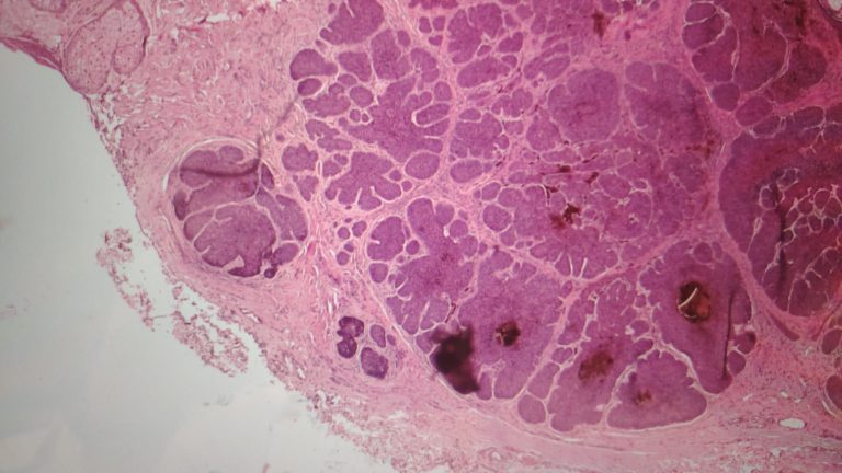
Detailed Process of Pathological Examination
The pathological examination process consists of several steps:
1.Sample Preparation
- The mole is sliced extremely thinly, almost transparent, for easier microscopic observation.
2.Staining Process
To make the transparent tissue and cells clearer, the pathology department uses dyes to stain the slices. For example, the now-common purple cell staining helps us identify features of basal cell carcinoma.
3.Microscopic Analysis
- Under the microscope, the nuclei of basal cell carcinoma cells typically arrange in a fence-like shape, which is a characteristic feature.
- If no cancer cell remnants are found in the slices surrounding the tumor, it can be confirmed that the cancerous tissue has been completely removed.
Post-surgery Follow-up Care
After the surgery, the patient only needs to return for regular follow-ups as directed by the doctor to remove stitches and monitor for signs of recurrence. Although the recurrence rate of basal cell carcinoma is low, close monitoring is still necessary.
Preventing Basal Cell Carcinoma
While the treatment for basal cell carcinoma is effective, prevention is always the best strategy.
1.Avoid Prolonged Sun Exposure
Reduce direct sunlight exposure during midday and regularly reapply sunscreen.
2.Be Aware of Skin Changes
Regularly check your skin and pay attention to changes in the shape and size of moles, spots, or wounds.
3.Healthy Diet and Lifestyle Habits
Strengthen your immune system by avoiding smoking and excessive drinking.
Hidden Content
Hidden content
Hidden Content
Hidden content